Leg Bones Diagram / 1000+ images about Bones in the Leg on Pinterest | Bone jewelry, Anatomy and Legs. Diagram and names of leg bones, diagram of foot and leg bones, diagram of leg bones, diagram of lower leg bones, diagram of the related posts of diagram of leg bones. High quality realistic skeleton legs. Your leg bones are the longest and strongest bones in your body. Pngtree offers bone diagram png and vector images, as well as transparant background bone diagram clipart images and psd files. Time to jump right into the biggest and strongest bones in the human body.
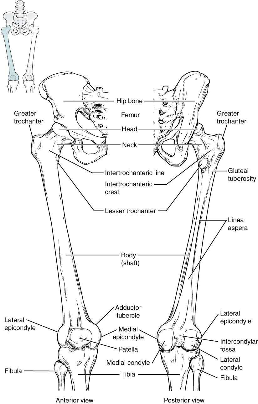
The foot bones shown in this diagram are the talus, navicular, cuneiform, cuboid, metatarsals and calcaneus. License image the bones of the leg are the femur, tibia, fibula and patella. At the microscopic level, this hard outer. The human leg, in the general word sense, is the entire lower limb of the human body, including the foot, thigh and even the hip or gluteal region. The foot bones shown in this diagram are the talus, navicular, cuneiform, cuboid.
Lupus and inexplicable connective tissue inflammation | Lupus, the Adventure Between the Lines from lupusadventurebetweenthelines.files.wordpress.com He leg's main function in the human is for locomotion and support of the rest of the body. Each leg is made up of four bones. The femur, or thighbone, is the longest and largest bone in the human body. Master leg and knee anatomy using our topic page. Time to jump right into the biggest and strongest bones in the human body. Diagram and names of leg bones, diagram of foot and leg bones, diagram of leg bones, diagram of lower leg bones, diagram of the related posts of diagram of leg bones. Includes leg (femur, tibia, patella, and fibula) and foot (tarsals and digits) bones. Use the leg bones diagrams to learn the names of the leg bones.
High resolution textures and displacement included.
The foot bones shown in this diagram are the talus, navicular, cuneiform, cuboid, metatarsals and calcaneus. Learn how to draw the femur, patella, tibia, and fibula in this lesson! Diagram of blood and nerve supply to bone. Its lower end helps create the knee joint. Click now to learn more about the bones, muscles, and soft tissues tibia: Pngtree offers bone diagram png and vector images, as well as transparant background bone diagram clipart images and psd files. At the same time, the bones and joints of the leg and foot must be strong enough to support the body's weight while remaining flexible enough for movement and balance. The human leg consists of 8 bones, 4 per leg. Each leg is made up of four bones. The foot bones shown in this diagram are the talus, navicular, cuneiform, cuboid, metatarsals. The musculoskeletal segment of the leg, including the foot bones (ankle, heel bone, toe bones), fibula and tibia, knee, femur and femoral neck, hip and sacrum as well as the third, fourth. Time to jump right into the biggest and strongest bones in the human body. Bones of the leg and foot, lower leg bone anatomy, leg bones anatomy, leg muscles, leg bones diagram, leg bone structure, leg anatomy muscles, parts of the lower leg.
At the same time, the bones and joints of the leg and foot must be strong enough to support the body's weight while remaining flexible enough for movement and balance. Use the leg bones diagrams to learn the names of the leg bones. The bones involved in it, however, are only the femur and the tibia, although the smaller bone of the leg, the fibula, is carried along in the movements of flexion, extension, and slight rotation that this joint. The foot bones shown in this diagram are the talus, navicular, cuneiform, cuboid, metatarsals. High resolution textures and displacement included.
Bones of the Lower Limb · Anatomy and Physiology from philschatz.com Learn how to draw the femur, patella, tibia, and fibula in this lesson! The human leg, in the general word sense, is the entire lower limb of the human body, including the foot, thigh and even the hip or gluteal region. Download the free graphic resources in the form of png, eps. Diagram of blood and nerve supply to bone. Leg bones (tibia and fibula). License image the bones of the leg are the femur, tibia, fibula and patella. The foot bones shown in this diagram are the talus, navicular, cuneiform, cuboid, metatarsals. Includes leg (femur, tibia, patella, and fibula) and foot (tarsals and digits) bones.
The femur, or thighbone, is the longest and largest bone in the human body.
Master leg and knee anatomy using our topic page. Quizzes on human skeletal system anatomy, bone anatomy, and bone markings. The bones of the leg are the femur, tibia, fibula and patella. The foot bones shown in this diagram are the talus, navicular, cuneiform, cuboid, metatarsals and calcaneus. At the same time, the bones and joints of the leg and foot must be strong enough to support the body's weight while remaining flexible enough for movement and balance. The human leg, in the general word sense, is the entire lower limb of the human body, including the foot, thigh and even the hip or gluteal region. These simple labelled diagrams of the bones of the lower legs and feet and the bones of the arms and hands this diagram shows the skeletal structure of the leg (anterior view) and foot (dorsal view). This bright worksheet helps your child bring these technical terms down to size. Visit kenhub for more skeletal system quizzes. He leg's main function in the human is for locomotion and support of the rest of the body. At the microscopic level, this hard outer. Health diagram bone skeleton leg knee science anchor chart human human body. When you stand or walk, all the weight of your upper body rests on them.
The foot bones shown in this diagram are the talus, navicular, cuneiform, cuboid, metatarsals and calcaneus. Leg bones (tibia and fibula). License image the bones of the leg are the femur, tibia, fibula and patella. High resolution textures and displacement included. Normal leg bones are relatively straight, but those affected by paget's disease are porous and figure 9.
Osteology Unit - M.Y. Online Portfolio from mkyousif17.weebly.com Its lower end helps create the knee joint. The largest and most medial leg bone, forming both the knee and ankle joints. Use the leg bones diagrams to learn the names of the leg bones. The foot bones shown in this diagram are the talus, navicular, cuneiform, cuboid. Diagram of blood and nerve supply to bone. He'll boost his body knowledge as he matches up the names of the bones with their proper places on the leg diagram. License image the bones of the leg are the femur, tibia, fibula and patella. High resolution textures and displacement included.
The human leg consists of 8 bones, 4 per leg.
The musculoskeletal segment of the leg, including the foot bones (ankle, heel bone, toe bones), fibula and tibia, knee, femur and femoral neck, hip and sacrum as well as the third, fourth. The human leg consists of 8 bones, 4 per leg. Bones of the leg and foot, lower leg bone anatomy, leg bones anatomy, leg muscles, leg bones diagram, leg bone structure, leg anatomy muscles, parts of the lower leg. Click now to learn more about the bones, muscles, and soft tissues tibia: The bones of the leg are the femur, tibia, fibula and patella. At the same time, the bones and joints of the leg and foot must be strong enough to support the body's weight while remaining flexible enough for movement and balance. Visit kenhub for more skeletal system quizzes. This bright worksheet helps your child bring these technical terms down to size. Health diagram bone skeleton leg knee science anchor chart human human body. The foot bones shown in this diagram are the talus, navicular, cuneiform, cuboid, metatarsals. He leg's main function in the human is for locomotion and support of the rest of the body. Quizzes on human skeletal system anatomy, bone anatomy, and bone markings. These simple labelled diagrams of the bones of the lower legs and feet and the bones of the arms and hands this diagram shows the skeletal structure of the leg (anterior view) and foot (dorsal view).
Share :
Post a Comment
for "Leg Bones Diagram / 1000+ images about Bones in the Leg on Pinterest | Bone jewelry, Anatomy and Legs"


Post a Comment for "Leg Bones Diagram / 1000+ images about Bones in the Leg on Pinterest | Bone jewelry, Anatomy and Legs"